Sensory adaptation: why your receptors go quiet when nothing changes
Meissner, Pacini, Merkel, Ruffini: why some receptors go quiet under constant pressure, and what that changes for your measurement in session.
The RNP training space, free for life
Get access to RNP training, no strings attached.
A hand resting on a table ends up barely feeling the table. This silence has a precise physiology, and it says a lot about what a sustained pressure sends to the nervous system.
Hello to you, the movement professional,
Put your hand flat on the table, right now. Stop moving and count slowly to ten. The table under your palm fades, gets blurry, almost absent. Slide one finger two millimeters: it comes back all at once, sharp, with its grain.
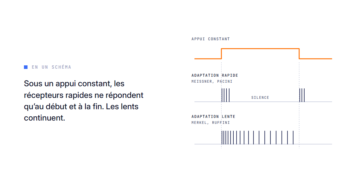
What you just felt is called sensory adaptation. A receptor exposed to a stimulus that no longer changes fires less and less. Some, like the Meissner and Pacini corpuscles, adapt quickly and only signal change; others, like the Merkel discs and the Ruffini endings, adapt slowly and keep signaling what lasts (Purves et al., 2004, ch. 8).
We came across this mechanism in our article on the tickle that disappears after thirty seconds of pressure. First-year physiology, you might say. Yet it affects your measurement: if you test a person right after a prolonged pressure, part of what you record comes from the state of their receptors at that very second.
Under your motionless palm, who went quiet, and who was still talking?
Fast-adapting and slow-adapting receptors: who codes what
Glabrous skin, that of the palm and the sole of the foot, carries four families of low-threshold mechanoreceptors (Purves et al., 2004, ch. 8; Enoka, 2008, ch. 7). The Meissner corpuscles, just under the epidermis, fire at the beginning and at the end of a contact, then go quiet during the sustained phase; they prefer slow vibrations, from 30 to 50 Hz, like those of a texture sliding under the finger. The Pacini corpuscles, deeper, adapt even faster and respond to fast vibrations, from 250 to 350 Hz. These are the so-called phasic receptors.
The Merkel discs and the Ruffini endings, for their part, keep firing as long as the stimulus lasts: the shape and edges of whatever is pressing for the former, skin stretch and its direction for the latter (Enoka, 2008, ch. 7). They are called tonic.
On your hand, after ten seconds, Meissner and Pacini had gone quiet while Merkel and Ruffini repeated the same information. The two millimeters of sliding woke the fast ones up.
A fast-adapting receptor signals a transition, never a state.
Holding an object and feeling it slip: why the body keeps both families
Take a glass. Motionless in your hand, it stays signaled by the slow receptors. If it starts to slip, you need to know quickly. Johansson and Westling (1987) recorded the tactile fibers of the fingers while volunteers lifted a small object between thumb and index finger: small slips excited these fibers, and the grip automatically tightened in about 74 milliseconds, roughly half the minimum delay of a voluntary correction.
Three types of units could signal these slips, and one of them belongs to the slow family: the two fast ones, plus the SA I (Merkel), all with strong dynamic sensitivity. Fast and slow describe a firing tendency, and the boxes in the table are less watertight than they seem.
For the hand, the synthesis by Johnson (2001) concludes that each of the four systems serves a distinct perceptual function. To simplify: with fast receptors only, the body would feel the glass arriving, then lose track of it while holding it.
And under the foot, which carries the body for a good part of the day, does the distribution look like that of the hand?
Mechanoreceptors of the foot sole: 71% fast-adapting units
Kennedy and Inglis (2002) recorded one by one the sensory fibers of the foot sole in 13 adults. Out of 104 units: 57% FA I, 14% FA II, 14% SA I and 15% SA II, that is 71% fast-adapting units. They were spread over the whole sole, with no concentration in the toes, and none fired spontaneously on an unloaded foot.
The hand follows another logic. Across 334 units, Johansson and Vallbo (1979) find a density that rises from the palm to the fingertip, from 58 to about 241 units per square centimeter according to their model. As for proportions, Purves puts forward for the hand about 40% Meissner, 25% Merkel, 20% Ruffini and 10 to 15% Pacini.
These figures hold for the hand, those of Kennedy and Inglis for the foot, and they are not interchangeable. The authors themselves note that plantar receptors "behave differently from those receptors found on the glabrous skin of the hand" (Kennedy and Inglis, 2002), and link this difference to the foot's role in balance.
A sole dominated by sensors of change should quickly lose track of a motionless pressure. That is our reading of the figures, which none of the cited studies measured as such.
The muscle spindle adapts too: dynamic response and static response
Proprioception follows the same logic. The muscle spindle, a stretch sensor placed in parallel with the muscle fibers, carries two endings. The primary one, connected to a Ia fiber, responds to length and above all to the speed of lengthening; the secondary one, connected to a group II fiber, informs mainly about length (Purves et al., 2004, ch. 8). In humans, Macefield and Knellwolf (2018) specify that both are sensitive to length as well as to velocity, with the primary keeping the greater dynamic sensitivity.
Unlike the skin sensors, the spindle receives its own motor command, the gamma motor neurons, which adjust its sensitivity (Enoka, 2008, ch. 7). The rest of the family is in our article on proprioceptive sensors.
One question remains that the hand experiment leaves open: did the fading come from the skin alone?
Sensory adaptation or habituation: the dishabituation test
Both lower a response, and they are often confused. Adaptation happens at the receptor, whose ending fires less. Habituation happens further along, in the central synapses; in Aplysia, Castellucci and Kandel (1974) tied it to a smaller release of neurotransmitter by the sensory neuron, the target neuron keeping its sensitivity.
To tell them apart, Rankin and colleagues (2009), who revised the definition of habituation, retain dishabituation. You present a new stimulus, on another area, then you return to the starting stimulus. If the response goes back up, the receptor was not the bottleneck, since nothing touched it in the meantime: the drop was habituation. If it stays low, adaptation, or a fatigue of the motor response, remains on the table.
Your table test probably mixes both, in a proportion it does not let you quantify.
What receptor adaptation changes for your session and your measurement
A motionless pressure held for a long time ends up speaking only to the slow receptors. Variation and movement wake up the fast ones, and under the foot they are the most numerous. Each receptor has its preferred stimulus, what we call its valence: choosing a stimulation means choosing who you are talking to. Whether a varied stimulus keeps adaptation at bay better, none of the studies cited here has tested.
The other consequence concerns measurement. Assessing a person's sensitivity, support or balance right after a sustained pressure amounts to measuring, in part, a receptor in the process of adapting, perhaps a central habituation. An immediate effect proves nothing; in our professions, what counts is the retest at a distance, and that is built through programming. Our grid says it in three steps: read where the body is in the dark, switch back on the missing input, make it last. Adaptation explains why the second step is often visible within the minute, and why the third is only judged at the retest.
We can be wrong, and it can be checked: the sensitivity to light touch of an area of the sole should drop right after thirty seconds of motionless pressure and return to its starting value at ten minutes. A drop still clear the next day, under the same conditions, would point to something other than short-term adaptation and habituation.
Claim | Level of evidence | Source |
|---|---|---|
71% fast units in the sole | Established, 104 units, 13 adults | Kennedy and Inglis |
Slip signaled by FA I, FA II and SA I | Established for the hand | Johansson and Westling |
Biased measurement right after a sustained pressure | Deduction, not quantified | The RNP reading |
What adaptation research has not settled yet
We do not know how long the receptors of the sole take to recover after a prolonged pressure in a standing person, and Kennedy and Inglis recorded an unloaded foot. We also do not know whether training modifies adaptation itself, nor how the share of the receptor and the share of the brain are distributed from one person to another.
Repeat the hand experiment tonight, then count how many of your session tests fall right after ten seconds of stillness.
Right after a sustained pressure, you are measuring a receptor that is recovering; only the next day do you measure what held.
👉 I want to intervene at the right place
Frequently asked questions
What is sensory adaptation?
The drop in firing of a receptor exposed to a stimulus that no longer changes. It happens in the sensor itself, before any central synapse.
What is the difference between phasic and tonic receptors?
Phasic receptors, which adapt quickly (Meissner, Pacini), fire at the beginning and at the end of a stimulus. Tonic receptors, which adapt slowly (Merkel, Ruffini), fire for as long as it lasts.
Does the sole of the foot have the same receptors as the hand?
The same families, in other proportions: 71% fast units in the sole (Kennedy and Inglis, 2002). The figures for the hand do not transfer to the foot.
How can you tell whether a drop in response comes from the receptor or from the brain?
Through dishabituation: a new stimulus, then a return to the starting stimulus. If the response comes back, the drop was central (Rankin et al., 2009).
By the LabO RNP team
LabO RNP is neither a medical nor a paramedical structure. Our approaches complement a care pathway, they never replace it.
References
Purves D, Augustine GJ, Fitzpatrick D, et al. Neuroscience, 3rd ed. Sunderland (MA): Sinauer Associates; 2004. Chapter 8 (somatosensory system).
Enoka RM. Neuromechanics of Human Movement, 4th ed. Champaign (IL): Human Kinetics; 2008. Chapter 7.
Johansson RS, Westling G. Signals in tactile afferents from the fingers eliciting adaptive motor responses during precision grip. Exp Brain Res. 1987;66(1):141-154. doi:10.1007/bf00236210. PMID 3582528.
Johnson KO. The roles and functions of cutaneous mechanoreceptors. Curr Opin Neurobiol. 2001;11(4):455-461. doi:10.1016/s0959-4388(00)00234-8. PMID 11502392.
Kennedy PM, Inglis JT. Distribution and behaviour of glabrous cutaneous receptors in the human foot sole. J Physiol. 2002;538(3):995-1002. doi:10.1113/jphysiol.2001.013087. PMID 11826182.
Johansson RS, Vallbo AB. Tactile sensibility in the human hand: relative and absolute densities of four types of mechanoreceptive units in glabrous skin. J Physiol. 1979;286:283-300. doi:10.1113/jphysiol.1979.sp012619. PMID 439026.
Macefield VG, Knellwolf TP. Functional properties of human muscle spindles. J Neurophysiol. 2018;120(2):452-467. doi:10.1152/jn.00071.2018. PMID 29668385.
Castellucci V, Kandel ER. A quantal analysis of the synaptic depression underlying habituation of the gill-withdrawal reflex in Aplysia. Proc Natl Acad Sci USA. 1974;71(12):5004-5008. doi:10.1073/pnas.71.12.5004. PMID 4373738.
Rankin CH, Abrams T, Barry RJ, et al. Habituation revisited: an updated and revised description of the behavioral characteristics of habituation. Neurobiol Learn Mem. 2009;92(2):135-138. doi:10.1016/j.nlm.2008.09.012. PMID 18854219.
Read next
Sensory Sensors - Proprioception
Learning pyramid and posturology: posture as a load-bearing level
Learning pyramid and posturology: posture at levels 3 and 4, its sensors, its link with attention and gaze, and the level of evidence.
Read the article Sensory Sensors - ProprioceptionThe sensory systems of the learning pyramid: why the body's senses come first
Sensory systems of the learning pyramid: tactile, vestibular and proprioceptive, habituation, interoception, and how to read them in the athlete.
Read the article Sensory Sensors - ProprioceptionLight Touch, Rhythmic Pressure, Deep Pressure: Why the Same Tactile Stimulation Doesn't Work for Everyone
Light touch, rhythmic or deep pressure, vibration: each tactile receptor has its preferred stimulus. Why the same gesture divides responses.
Read the articleReady to read the loop before the movement?
The RNP system is meant to be read, targeted and tested from your next assessment. Start with the member space, for free.