Light Touch, Rhythmic Pressure, Deep Pressure: Why the Same Tactile Stimulation Doesn't Work for Everyone
Light touch, rhythmic or deep pressure, vibration: each tactile receptor has its preferred stimulus. Why the same gesture divides responses.
The RNP training space, free for life
Get access to RNP training, no strings attached.
Two people, an identical gesture under the foot, and two responses that have nothing in common. The physiology of tactile receptors explains a good part of the gap; the rest is decided before touching.
Hello to you, the movement professional,
The scene comes back often in sessions. Two people receive exactly the same stimulation under the foot, on the same area and for the same duration. The first stands up straighter, her stance widens, and she says she feels better grounded. The second gets off the table shrugging: she feels nothing in particular.
A good part of the gap comes from the receptors. The sole houses several families of tactile receptors, and each responds preferentially to one type of contact: light touch speaks mostly to the fast receptors, sustained pressure to the slow receptors, and vibration to one or the other depending on its frequency. A stimulation therefore chooses its interlocutors. If it misses the ones the person needed, little happens, and the technique is not at fault.
We often hear that the second person is "less receptive". Perhaps, but you can only say so after looking at whom the stimulation was addressing. As for the first, her visible effect within the minute says nothing about how long it lasts: we showed this with the tickle that disappears after thirty seconds of pressure.
The families of tactile stimulation you meet in the field
In practice, skin stimulations come down to a handful of families: light touch, light and mobile, with fingertips or a brush; slow stroking, broader; rhythmic pressure, which presses and releases; long, deep pressure, which settles in; vibration; and texture, when the foot meets a granular, spiked or soft surface. We use them all, like many colleagues.
From a distance, it is always hands on a foot; the skin, though, tells them apart, because each gesture deforms it in its own way. Light touch slides across the surface, deep pressure compresses and stays, vibration shakes the tissues tens or hundreds of times per second.
You still need to know who is listening, under the sole.
Fast and slow tactile receptors: what the textbooks describe
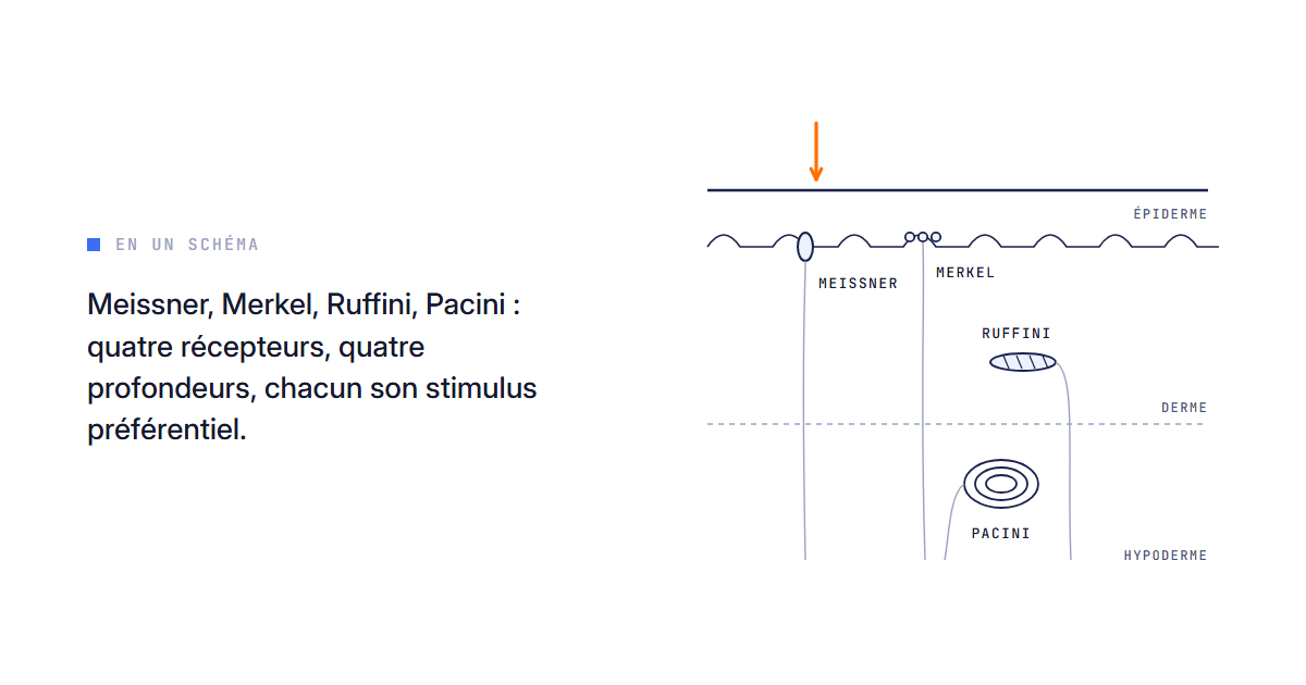
The sole of the foot is glabrous skin, without hair, like the palm. It houses four families of low-threshold cutaneous mechanoreceptors, sensitive to small deformations. Two adapt quickly: they fire when contact begins, changes or stops, then fall silent if it stays still. These are the Meissner and Pacini corpuscles. The other two, Merkel discs and Ruffini endings, adapt slowly and signal a pressure or a stretch for as long as it lasts.
The textbooks give them clear specialties. Meissner signals slip and fine texture, and prefers low-frequency vibrations, a few tens of hertz. Pacini, deeper, picks up very rapid pressure changes and high-frequency vibrations, several hundred hertz. Merkel codes shape and edges, Ruffini the stretch of the skin and its direction (Purves et al., 2004, ch. 8; Enoka, 2008, ch. 7). By textbook logic, light touch and rhythmic pressure, made of changes, mostly engage the fast receptors; under a sustained pressure, the fast ones fall silent and the slow ones carry the signal.
Under the foot, the distribution has been measured fiber by fiber. Kennedy and Inglis (2002) found a majority of fast receptors there. Strzalkowski et al. (2018) have since gathered in a review 364 afferents from the sole: 51% of the type associated with Meissner, 12% of the Pacini type, 17% of the Merkel type and 20% of the Ruffini type. Their firing thresholds are about ten times higher than in the hand, and density varies from one zone to another: higher at the toes and on the outer edge, lower at the heel and on the inner edge. In short, a sensor built to carry body weight, less fine than the hand.
Yet one family is missing from this inventory.
Slow stroking and C-tactile afferents
In hairy skin, such as the forearm or the back, another system responds to light touch: slow, unmyelinated fibers called C-tactile afferents. Olausson et al. (2002) studied them in a person deprived of her large myelinated fibers. Their activation produced a faint sensation of pleasant touch, and functional MRI showed the insular region activated, without the primary and secondary somatosensory areas. The authors see in this a possible basis for emotional responses to skin-to-skin contact. Löken et al. (2009) then showed that when a soft brush passes over, these fibers fire most at intermediate stroking speeds, the ones participants rated as most pleasant; the myelinated fibers did not follow this profile.
A word of caution for our subject: these fibers have been described mostly in hairy skin, and the sole is glabrous skin. Transposing these results to the foot would go further than the studies. Deep pressure, for its part, was the subject of our article on touch and sleep.
The valence principle: each receptor has its preferred stimulus
Put end to end, these facts outline a principle. A review devoted to the sole sums it up in one line, referring to Johnson (2001): "each cutaneous afferent class preferentially encodes distinct tactile stimuli" (Strzalkowski et al., 2018). Abraira and Ginty (2013) speak of tuning properties specific to each receptor subtype.
At LabO RNP, we call this valence (the word sounds a bit like chemistry, and we own it): the type of stimulus a receptor responds to best. We have drawn a valence grid from it, which we teach in training, always with the reading that goes with it. The principle, which underpins the sensory specificity of stimulation, fits in one sentence: choosing a stimulation means choosing which receptors you are addressing.
Two nuances keep it from becoming a dogma. Preferential means preferential: a real touch deforms the skin in several ways at once and recruits a mixture of receptors. And thresholds shift with the zone, the state of the skin, the load the foot carries at the moment of contact. Valence guides a choice, without guaranteeing a result.
So where does the gap between our two people come from?
Same stimulation, two opposite responses
Same gesture, nearly the same receptors engaged; the need, though, differed. Our field reading, presented as such: in the first person, the stimulation landed on an under-used input and brought to the control of stance the information that was missing. In the second, it spoke to receptors that were already well served. When a stimulation gives nothing, it often did not speak to the receptors that person needed.
This reading has limits, so let us state them. Attention, expectation, fatigue or the way of asking "do you feel a difference?" produce the same contrast. A gap between two people remains a correlation, and nothing proves that valence explains it in a given case. We told a similar situation in two identical sessions with opposite results.
And what if the first person had simply had a good time?
The immediate effect settles nothing
We all want to judge a stimulation by the visible straightening within the minute. Yet a tactile stimulation changes the state of the system for a short time: fast receptors adapt, the system habituates, then the response comes back on its own (Rankin et al., 2009). The immediate effect in the first person does not guarantee it will last, and the absence of an effect in the second does not condemn the stimulation.
In our professions, what counts is what holds over time, measured at the retest and built through programming.
A verifiable prediction: if valence played no role, two stimulations with opposite valences, in the same person, would give indistinguishable results at a delayed retest. If that is what we observe repeatedly, our grid loses its reason to exist.
Read before stimulating
Our way of working follows the image of the body in the dark: read where it is dark, switch the missing input back on, make it hold. Without a reading, you choose your stimulation by habit or by taste, and you credit to the technique what belonged to the receptor. With a reading, valence becomes a selection criterion, and the retest tells you whether the choice was right.
Claim | Level of evidence |
|---|---|
Fast and slow receptors, each with its specialty | Established (Purves; Enoka; Johnson) |
Distribution and thresholds of sole receptors | Established by microneurography (Kennedy and Inglis; Strzalkowski) |
C-tactile afferents and slow stroking | Established in hairy skin (Olausson; Löken), not established for the sole |
Same gesture, opposite responses depending on the person's need | Field reading, not tested under controlled conditions |
What physiology does not yet settle
No field measurement today reliably identifies the family of receptors at fault in a given person. We also do not know whether C-tactile afferents play a role under the foot. And, to our knowledge, no study has compared stimulations chosen after a reading with randomly drawn stimulations, with a delayed retest.
The next time a stimulation gives nothing, before changing technique, ask yourself whom it was speaking to.
A stimulation is worth what the reading that chose it is worth.
👉 I want to intervene in the right place
Frequently asked questions
Which tactile receptors respond to light touch?
Mostly the rapidly adapting receptors, first and foremost the Meissner corpuscles, which signal slip. They fall silent as soon as the contact stays still.
What is the difference between rhythmic pressure and deep pressure for receptors?
Rhythmic pressure restarts the fast receptors at every change. Under sustained pressure, they adapt, and the slow receptors, Merkel and Ruffini, keep signaling.
Is an immediately visible effect enough to validate a stimulation?
No, adaptation and habituation produce quick effects that fade. Only a delayed retest tells you whether anything held.
By the LabO RNP team
LabO RNP is neither a medical nor a paramedical organization. Our approaches complement a care pathway and never replace it.
References
Purves D, Augustine GJ, Fitzpatrick D, et al. Neuroscience, 3rd ed. Sunderland (MA): Sinauer Associates; 2004. Chapter 8 (somatosensory system).
Enoka RM. Neuromechanics of Human Movement, 4th ed. Champaign (IL): Human Kinetics; 2008. Chapter 7.
Kennedy PM, Inglis JT. Distribution and behaviour of glabrous cutaneous receptors in the human foot sole. J Physiol. 2002;538(3):995-1002. PMID 11826182.
Strzalkowski NDJ, Peters RM, Inglis JT, Bent LR. Cutaneous afferent innervation of the human foot sole: what can we learn from single-unit recordings? J Neurophysiol. 2018;120(3):1233-1246. doi:10.1152/jn.00848.2017. PMID 29873612.
Johnson KO. The roles and functions of cutaneous mechanoreceptors. Curr Opin Neurobiol. 2001;11(4):455-461. doi:10.1016/s0959-4388(00)00234-8. PMID 11502392.
Abraira VE, Ginty DD. The sensory neurons of touch. Neuron. 2013;79(4):618-639. doi:10.1016/j.neuron.2013.07.051. PMID 23972592.
Olausson H, Lamarre Y, Backlund H, et al. Unmyelinated tactile afferents signal touch and project to insular cortex. Nat Neurosci. 2002;5(9):900-904. doi:10.1038/nn896. PMID 12145636.
Löken LS, Wessberg J, Morrison I, McGlone F, Olausson H. Coding of pleasant touch by unmyelinated afferents in humans. Nat Neurosci. 2009;12(5):547-548. doi:10.1038/nn.2312. PMID 19363489.
Rankin CH, Abrams T, Barry RJ, et al. Habituation revisited: an updated and revised description of the behavioral characteristics of habituation. Neurobiol Learn Mem. 2009;92(2):135-138. doi:10.1016/j.nlm.2008.09.012. PMID 18854219.
Read next
Sensory Sensors - Proprioception
Learning pyramid and posturology: posture as a load-bearing level
Learning pyramid and posturology: posture at levels 3 and 4, its sensors, its link with attention and gaze, and the level of evidence.
Read the article Sensory Sensors - ProprioceptionThe sensory systems of the learning pyramid: why the body's senses come first
Sensory systems of the learning pyramid: tactile, vestibular and proprioceptive, habituation, interoception, and how to read them in the athlete.
Read the article Sensory Sensors - ProprioceptionSensory adaptation: why your receptors go quiet when nothing changes
Meissner, Pacini, Merkel, Ruffini: why some receptors go quiet under constant pressure, and what that changes for your measurement in session.
Read the articleReady to read the loop before the movement?
The RNP system is meant to be read, targeted and tested from your next assessment. Start with the member space, for free.